

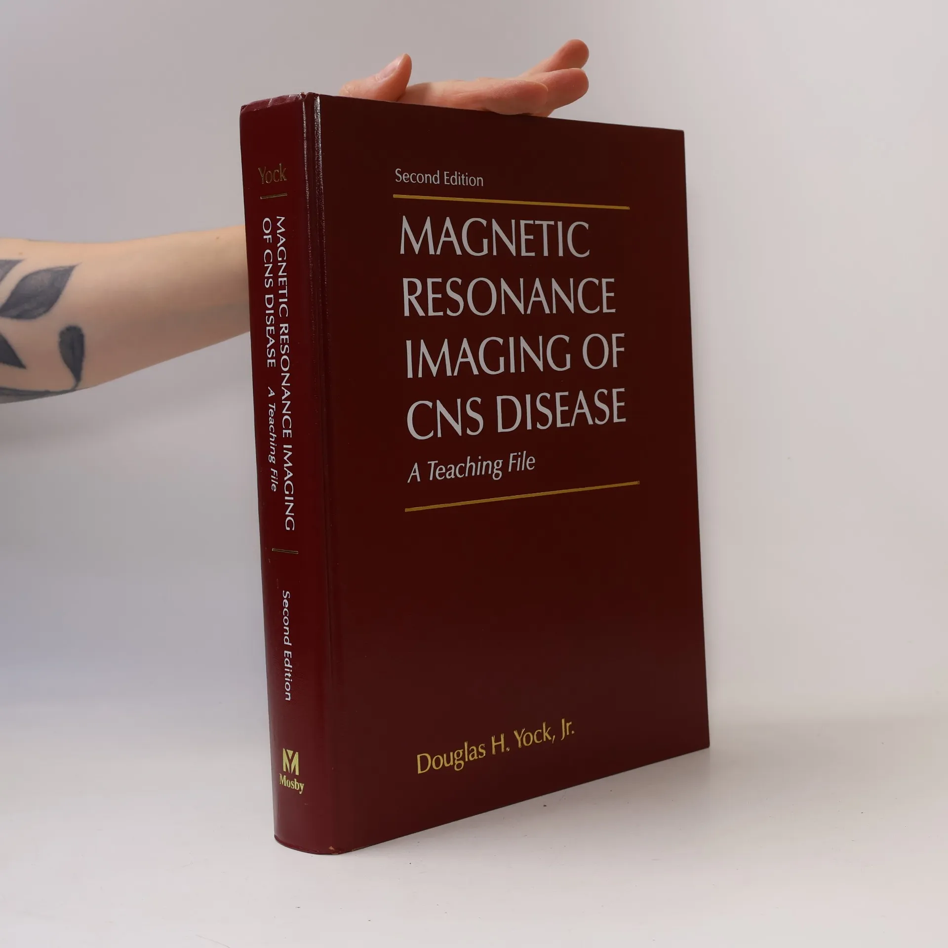


Magnetic Resonance Imaging of CNS Disease
A Teaching File - Second Edition
Autoři
Více o knize
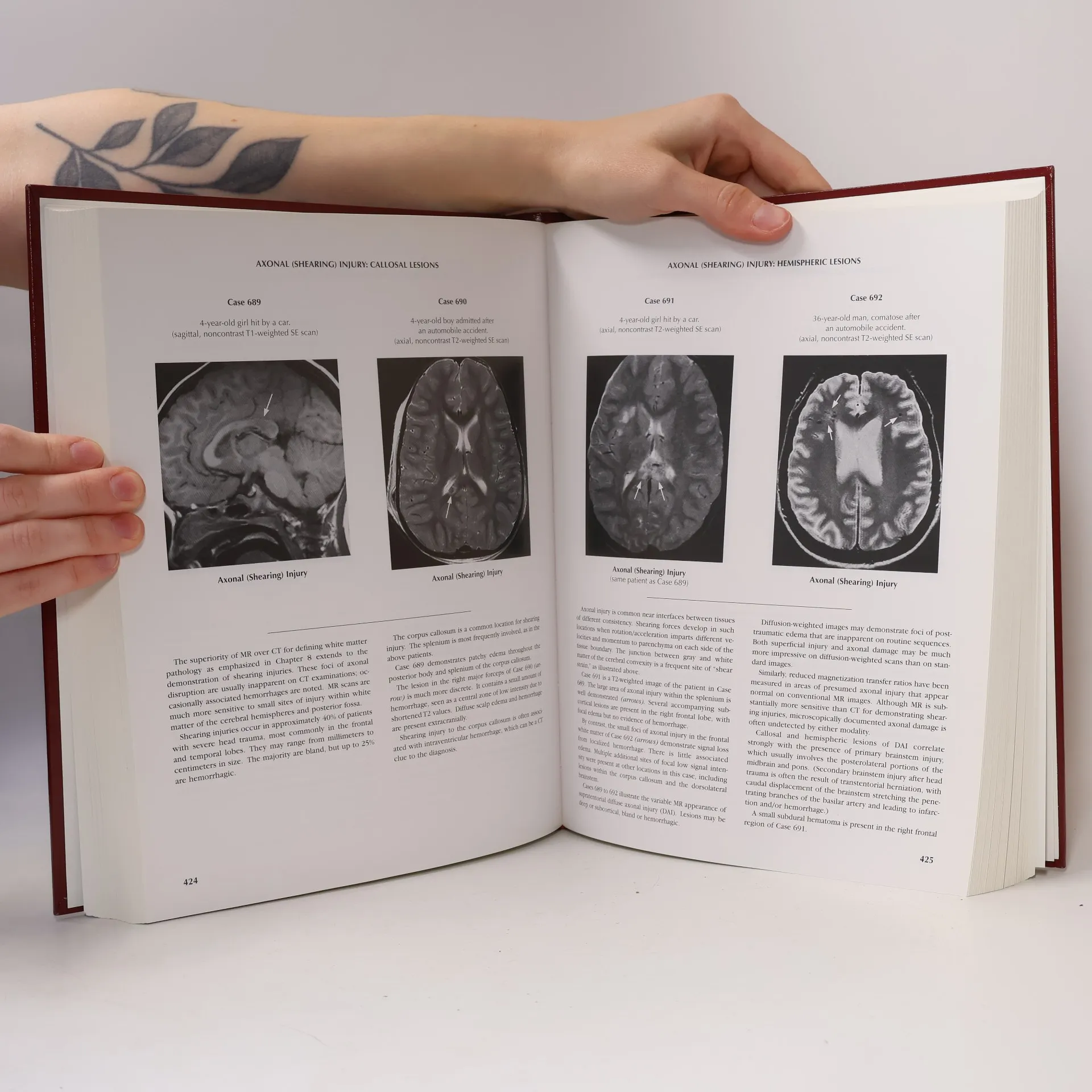
The thoroughly updated second edition of this user-friendly resource presents over 1200 carefully selected cases that cover the entire spectrum of CNS diseases affecting the head and spine, including newly recognized disorders like posterior reversible edema syndrome, spinal cord herniation, and dysembryoplastic neuroepithelial tumor. With over 1350 illustrations of neuropathology, it showcases both typical and unusual appearances. Hundreds of cross-references link cases to highlight similarities, distinctions, and variations among conditions. Consistent labeling of images allows for quick scanning of individual topics, and side-by-side comparisons illustrate potential similarities among different pathologies. The resource emphasizes the interpretation of MR scans as encountered in practice and includes over 150 illustrated differential diagnoses. It features a significantly expanded pictorial index that illustrates diagnostic possibilities within key MR finding patterns. Additionally, it presents the latest technical advances, such as the use of FLAIR sequences and improved MR angiography, along with the growing contributions of diffusion-weighted images and proton spectroscopy. The edition incorporates over 600 new cases, enhancing its comprehensive nature.
Nákup knihy
Magnetic Resonance Imaging of CNS Disease, Douglas H. Yock Jr.
- Jazyk

- Rok vydání
- 2002
- Vazba
- (pevná),
- Stav knihy
- Dobrá
- Cena
- 4 849 Kč
Doručení
Platební metody
Nikdo zatím neohodnotil.
- Titul
- Magnetic Resonance Imaging of CNS Disease
- Podtitul
- A Teaching File - Second Edition
- Jazyk
- anglicky
- Autoři
- Douglas H. Yock Jr.
- Vydavatel
- Mosby
- Rok vydání
- 2002
- Vazba
- pevná
- Počet stran
- 842
- ISBN10
- 0323011721
- ISBN13
- 9780323011723
- Série
- Kategorie
- Anotace
- The thoroughly updated second edition of this user-friendly resource presents over 1200 carefully selected cases that cover the entire spectrum of CNS diseases affecting the head and spine, including newly recognized disorders like posterior reversible edema syndrome, spinal cord herniation, and dysembryoplastic neuroepithelial tumor. With over 1350 illustrations of neuropathology, it showcases both typical and unusual appearances. Hundreds of cross-references link cases to highlight similarities, distinctions, and variations among conditions. Consistent labeling of images allows for quick scanning of individual topics, and side-by-side comparisons illustrate potential similarities among different pathologies. The resource emphasizes the interpretation of MR scans as encountered in practice and includes over 150 illustrated differential diagnoses. It features a significantly expanded pictorial index that illustrates diagnostic possibilities within key MR finding patterns. Additionally, it presents the latest technical advances, such as the use of FLAIR sequences and improved MR angiography, along with the growing contributions of diffusion-weighted images and proton spectroscopy. The edition incorporates over 600 new cases, enhancing its comprehensive nature.