Mimořádně instruktivní celobarevná publikace obsahuje na 550 vyobrazení, najdete v ní typické nálezy a důležité poučky, které umožní rychlou orientaci v sonografickém snímku i bez předchozích hlubších zkušeností. V knize naleznete vyobrazení všech sonograficky vyšetřovaných oblastí lidského těla. Publikace je vhodným doplňkem k učebnici sonografie.
Berthold Block Knihy




Abdominal ultrasound
- 292 stránek
- 11 hodin čtení
Designed to be kept close at hand during an actual ultrasound examination, Abdominal Ultrasound: Step by Step, second edition, provides the tools, techniques and training to increase your knowledge and confidence in interpreting ultrasound findings. Its clear, systematic approach shows you how to recognize all important ultrasound phenomena (especially misleading artifacts), locate and delineate the upper abdominal organs, explain suspicious findings, apply clinical correlations, and easily distinguish between normal and abnormal images. This second edition includes the new Sono Consultant, a
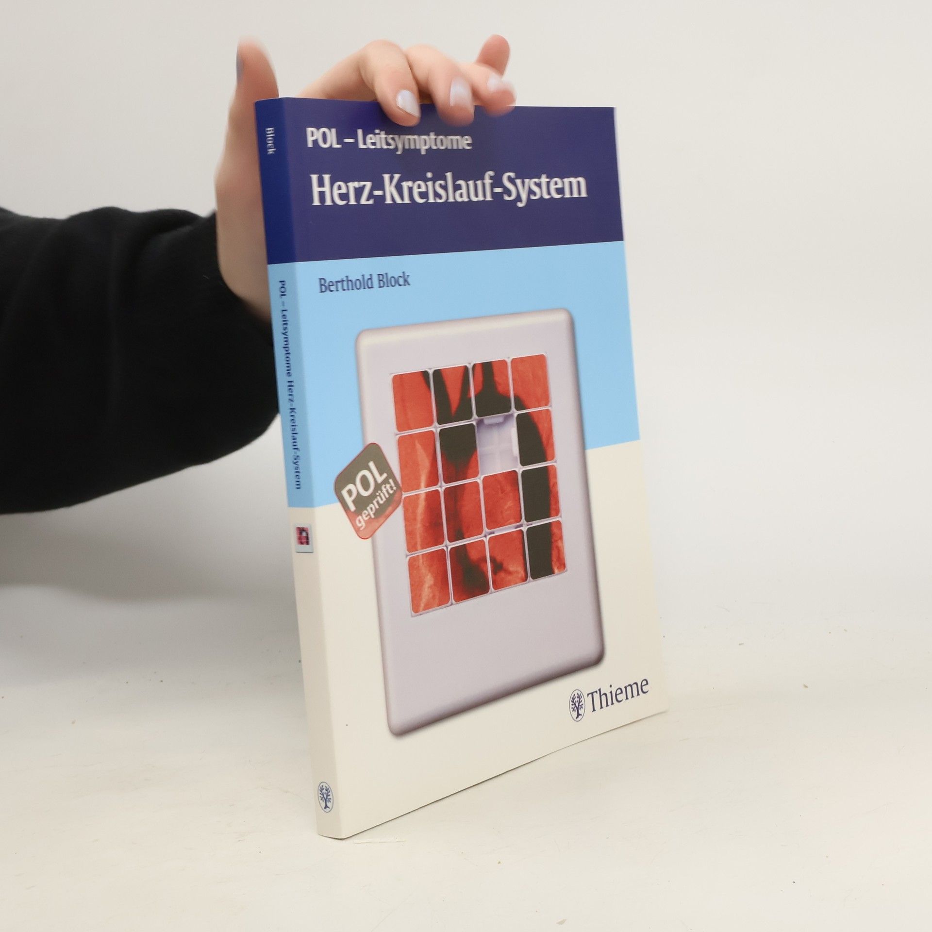
Mit POL zu mehr Praxisnähe - wie von der neuen AO gefordert! Die neue Approbationsordnung verlangt praxisnahes, fächerübergreifendes Lernen durch Methoden wie Bedside-Teaching, POL-Kurse und Fallbesprechungen. Bei Patienten mit Luftnot oder Schluckbeschwerden ist problemorientiertes Lernen (POL) besonders hilfreich. POL ist eine bewährte Methode, die neben klassischem Wissenserwerb auch die Entwicklung eigener Problemlösungsstrategien fördert. In Kleingruppen erarbeiten die Studierenden Lernziele zu klinischen Fragestellungen. Die neue Reihe stellt das Vorgehen bei wichtigen Leitsymptomen anhand des klassischen POL-Werkzeugs – den „7 steps“ – dar: Begriffe klären, Problem erkennen, Grundlagen rekapitulieren, mögliche Ursachen kennen, Problem schrittweise lösen, Anamneseerhebung, weitergehende Diagnostik, Diagnose sichern und Therapie einleiten. Diese Begleitliteratur ist optimal auf die Anforderungen der neuen Approbationsordnung zugeschnitten und bietet praxisnahe Ergänzungen durch komplexe Kasuistiken. Sie enthält typische Befunde ausgewählter Erkrankungen und einen „Serviceteil“ mit Informationen zur körperlichen Untersuchung und apparativen Diagnostik. Die wichtigsten Leitsymptome bei Erkrankungen des Herz-Kreislauf-Systems werden praxisnah und fächerübergreifend aufbereitet.
Dr. Blocks Patiententypologie
- 207 stránek
- 8 hodin čtení
Der Kraftmensch, der Tabletten-Freak oder der Patient, der auch bei seiner Krankheit mit der Mode geht: Auf den ersten Blick präsentiert uns Berthold Block ein Panoptikum skurriler Typen. Wer jedoch genauer hinsieht, erkennt sich selbst in seiner Rolle als verunsicherter Patient. Die pointierten Portraits sind nicht nur urkomisch und tiefsinnig, sie entlarven auch den Geist unseres Gesundheitssystems, der so manche Schrulligkeit geradezu heraufbeschwört.
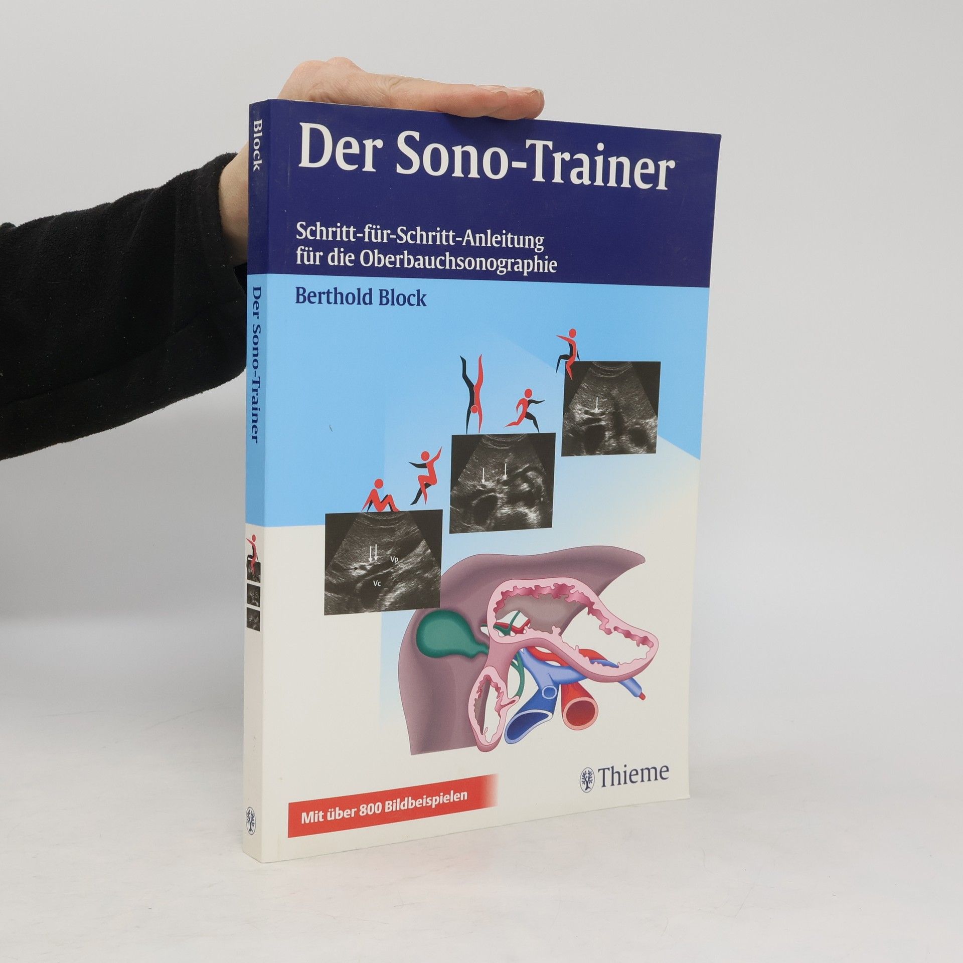
Der Sono-Trainer
Schritt-für-Schritt-Anleitungen für die Oberbauchsonografie
Der Schritt-für-Schritt-Selbstlernkurs für Ultraschall-Einsteiger bietet praxisorientierte Lerneinheiten mit über 700 Sonobildern und 250 Zeichnungen. Leser lernen, wichtige Ultraschallphänomene zu erkennen und zu interpretieren. Der digitale Zugang zur Wissensplattform eRef ermöglicht jederzeit Zugriff auf zusätzliche Inhalte.
Ultrasonografia jest doskonałym narzędziem w rękach doświadczonego lekarza. Ale od czegoś trzeba zacząć! I to jest właśnie książka na dobry początek nauki. W III wydaniu uznanego podręcznego atlasu przeredagowano i uzupełniono rozdziały dotyczące pęcherzyka żółciowego, śledziony i nerek. Poszerzono również rozdziały dotyczące gruczołu krokowego i macicy. W publikacji można znaleźć m.in.: 308 dokładnie oznakowanych i opisanych par rycin i zdjęć; standardowe płaszczyzny obrazowania poszczególnych narządów; topografię anatomicznych organów i struktur we wszystkich trzech płaszczyznach obrazowania. Publikacja jest adresowana do studentów medycyny, lekarzy radiologów, rezydentów i techników radiologii.
Facharztprüfung innere Medizin
- 498 stránek
- 18 hodin čtení
Facharztprüfung in Sicht? Verinnerlichen Sie das gesicherte Wissen aus allen Themengebieten der Inneren Medizin spielerisch durch Frage-Antwort-Kombinationen. So bleibt der Stoff im Kopf und Motivationstiefs haben keine Chance. Anhand fall- und problemorientierter Fragen lernen Sie, wie Sie Fakten richtig bewerten und klinische Probleme lösen. Ergänzt durch Wissenswertes zu Antragstellung, Ablauf der Prüfung und Tipps für einen souveränen Auftritt, bietet dieser Titel das Rundum-Sorglos-Paket für eine effektive Prüfungsvorbereitung. Plus: Das gute Gefühl, bestens vorbereitet in die Prüfung zu gehen.
Der Sono-Trainer
- 252 stránek
- 9 hodin čtení
Der Gastroskopie-Trainer
- 193 stránek
- 7 hodin čtení
Bedienen des Gerätes- Gerätetechnik und Handhabung des Endoskopes leicht verständlich dargestellt- 3D-Graphiken und Positionsskizzen erleichtern auf geniale Weise die räumlicheOrientierungUntersuchen- Checklisten und tabellarische Übersichten zu Untersuchungsvorbereitungen, Begleitmedikation, Komplikationen und Risiken etc.- Schritt-für-Schritt-Anleitungen zeigen den gesamten Untersuchungsablauf- das ganze Spektrum der normalen und pathologischen Befunde in über 700 Abbildungen- endoskopische Kriterien und die wichtigsten Differenzialdiagnosen aller Krankheitsbilder- Sicherheit durch Referenzbilder, Vergleichsabbildungen zur Befundvariabilität und Bilderserien- Musterbefunde und Formulierungshilfen zur korrekten DokumentationTherapierenFotoserien zu allen wichtigen invasiven endoskopische Therapie von Blutungen, Anlegen von PEG und Duodenalsonden, Fremdkörperentfernung etc.Plus- immer nur eine Lerneinheit pro Seite- Großzügiges Layout im großen Atlasformat
