Torsten B. Möller Knihy

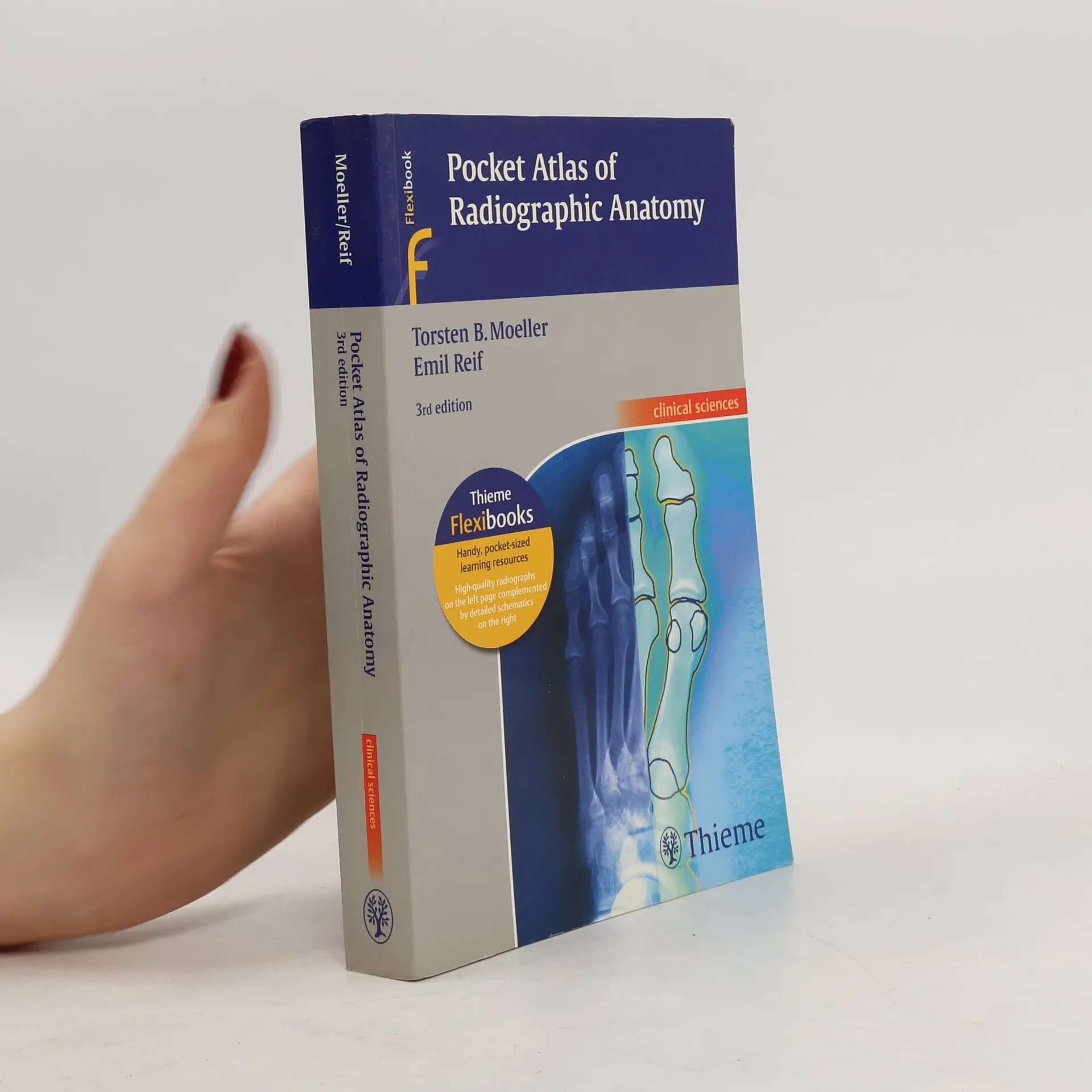

Flexibooks: Pocket Atlas of Radiographic Anatomy
Clinical Sciences - 3rd Edition
- 388 stránek
- 14 hodin čtení
Third edition of the classic pocket guide to basic radiographic anatomyIn this easily accessible pocket atlas, two expert radiologists present the normal radiographic anatomy readers need in order to interpret conventional diagnostic radiographs. Each practical, two-page unit displays a standard radiograph of a different projection on the left-hand side supplemented by a detailed, clearly labeled schematic drawing on the opposing page. The consistent, user-friendly format facilitates easy identification and rapid review of key anatomic information. Features:177 radiographic studies provide multiple views of every basic anatomic structure High-resolution radiographs appear beside explanatory drawings to aid comprehension Seven examinations new to this edition cover a trans-scapular „Y“ view of the shoulder; 45° external and internal rotation views of the knee; and moreAn ideal reference for anyone involved in the interpretation of commonly performed radiographic studies, the third edition of Pocket Atlas of Radiographic Anatomy is an especially valuable tool not only for medical students and radiology residents, but also for radiological technologists.
MRI parameters and positioning
- 341 stránek
- 12 hodin čtení
Packed with information on the practical aspects of MRI, this user-friendly text covers everything from advice on optimal positioning of patients to recommendations for setting the appropriate scanning parameters. Each consistently organized chapter follows the chronology of a standard procedure - the authors present essential information on preparation and necessary materials first. Then they skilfully guide the readers through special considerations in positioning and coil selection, protocols for conducting the exam, examples of various sequences, and possible modifications. Numerous tips, tricks, and pointers explain how to avoid potential complications. Highlights of the second edition: 340 high-quality MRI scans and anatomical drawings New and expanded sections on MR angiography of pulmonary arteries and pelvic and leg vessels; the CARE Bolus Technique; whole-body MRI; and more Information on the latest protocols for MR urography, cholangiography, and colonography Consistent chapter structure for maximum accessibility on the job and at the MRI workstation Each section contains plenty of space on each page for personal notes A guide to the most important MRI studies, the second edition of MRI Parameters and Positioning is an indispensable companion for all radiologists, radiology residents, and radiologic technologists.
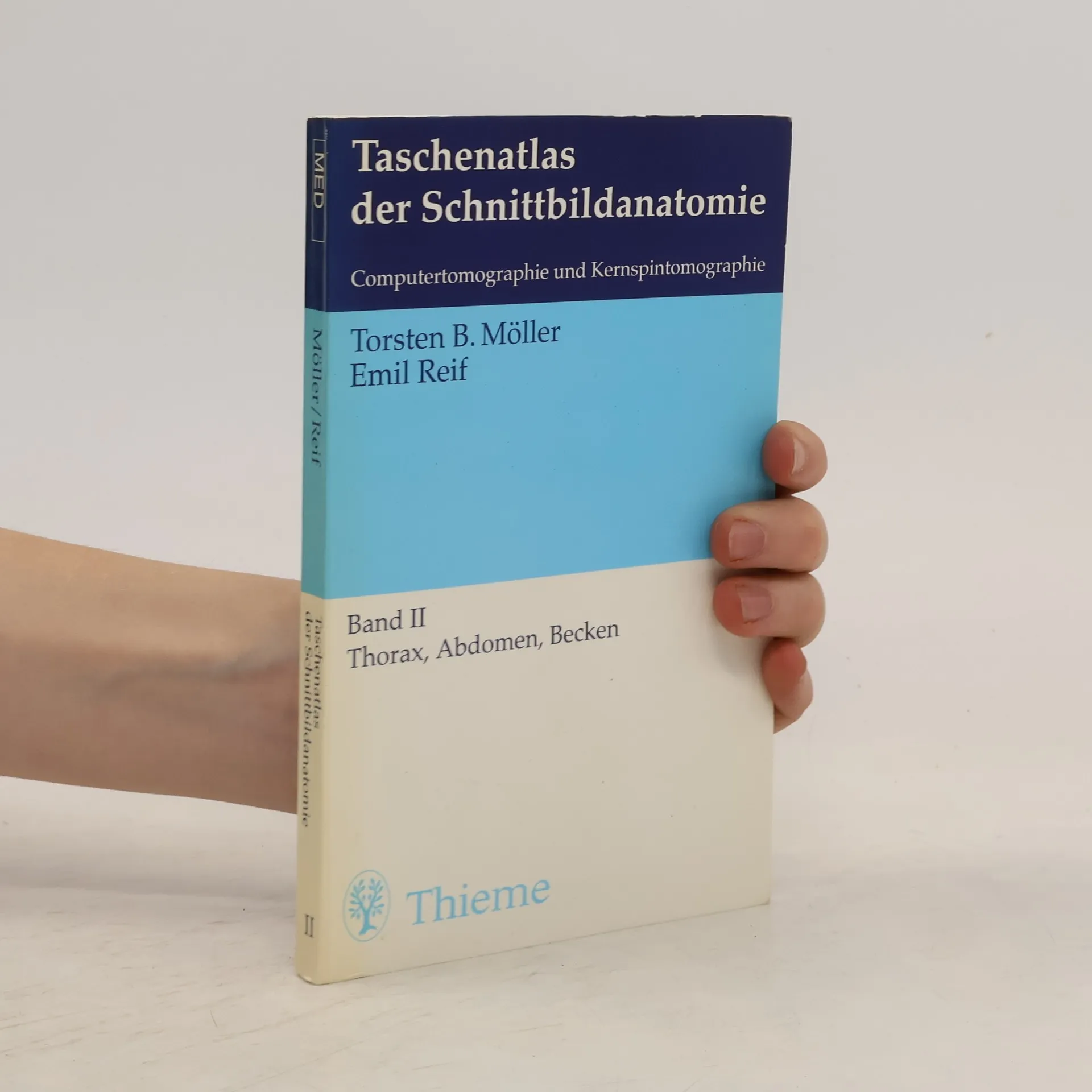
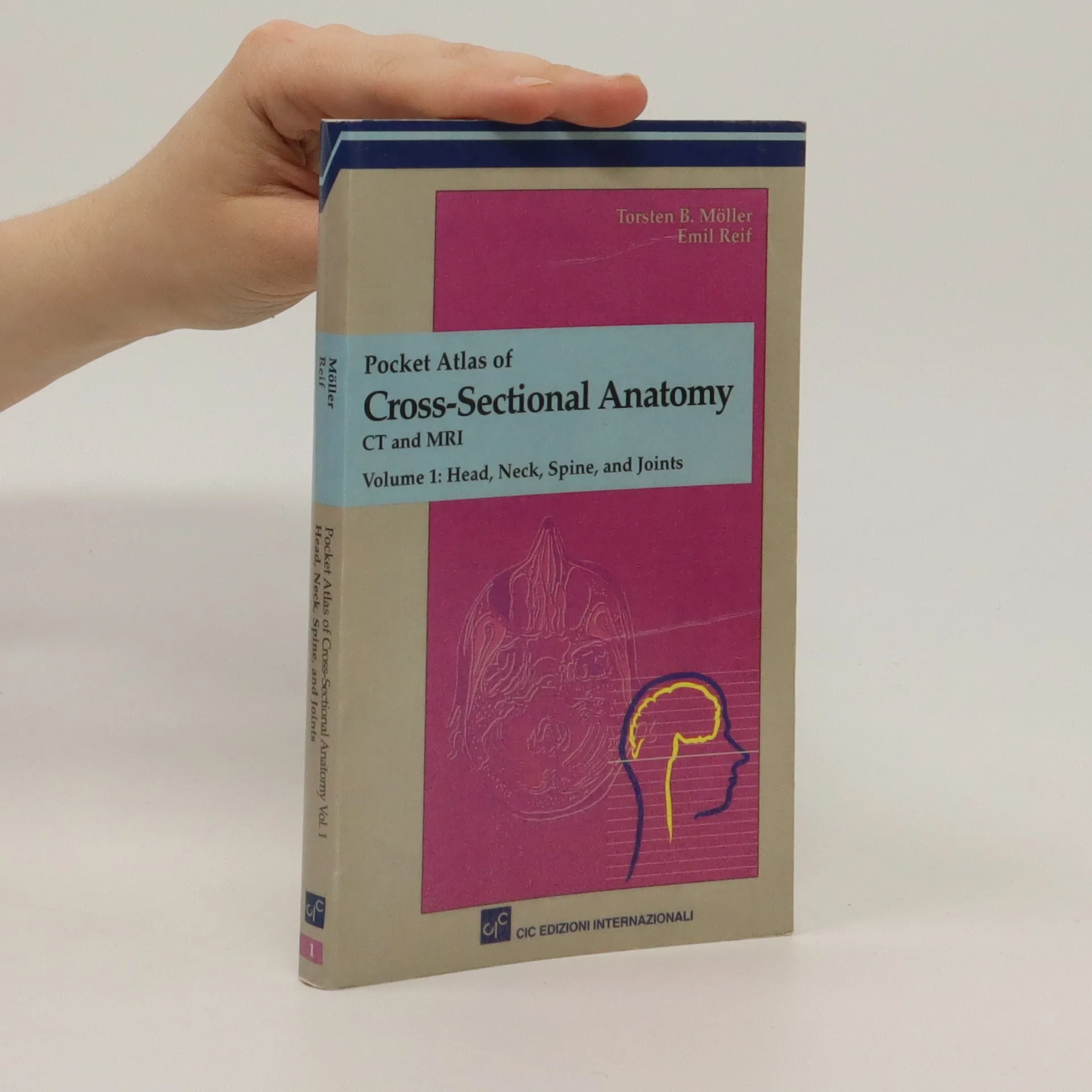
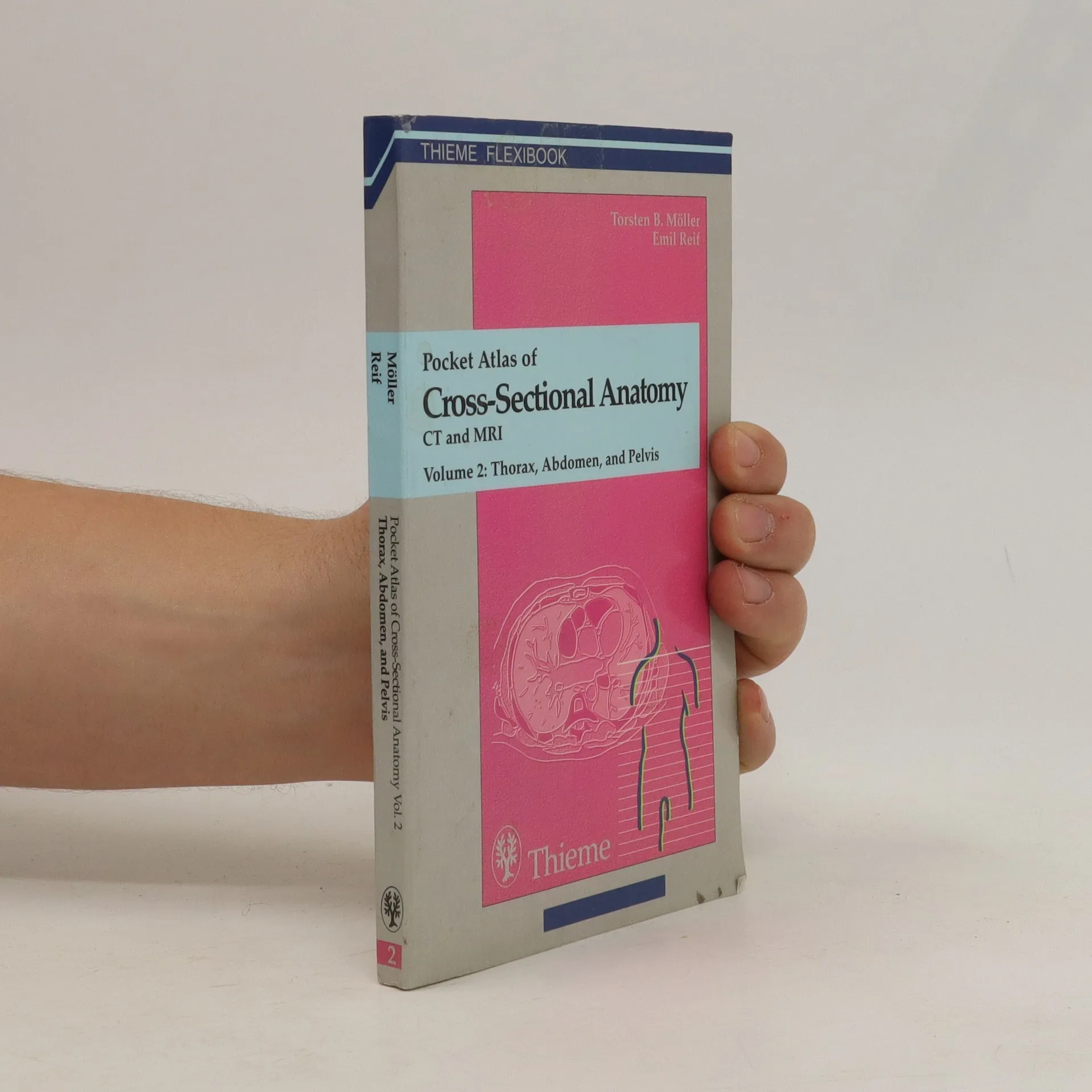
Pocket Atlas of Cross Sectional Anatomy
- 246 stránek
- 9 hodin čtení
The second of a two volume set which describes the anatomical details visualized in diagnostic tomography. As a comprehensive reference it is an aid when interpreting images for anatomic structures presented in representative cross-sectional CT and MRI images.
Thieme Flexibook: Normal Findings in CT and MRI
- 250 stránek
- 9 hodin čtení
The key for any beginning radiologist who wishes to recognize pathological findings is to first acquire an ability to distinguish them from normal ones. This outstanding guide gives beginning radiologists the tools they need to systematically approach and recognize normal MR and CT images. Highlights include: Reference-quality images from the author's own teaching files show all standard normal findings as seen in CT and MRI Checklists in each section offer the reader a systematic way to approach the images Thorough guidelines to help beginning radiologists dictate their reports Lists of standard measurements and tips for ruling out pathology
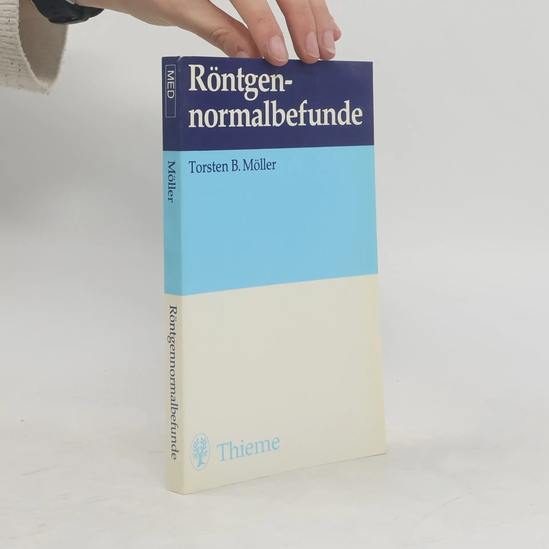
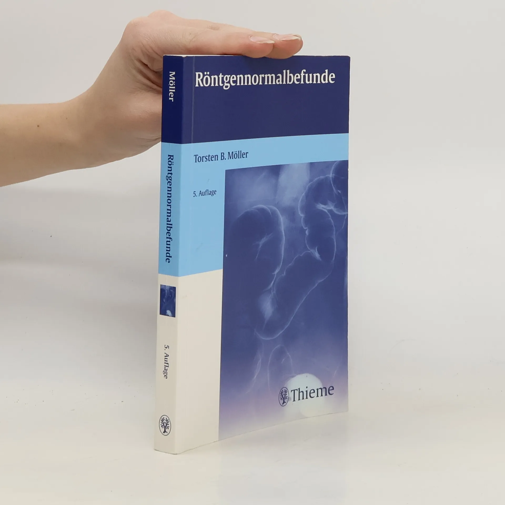
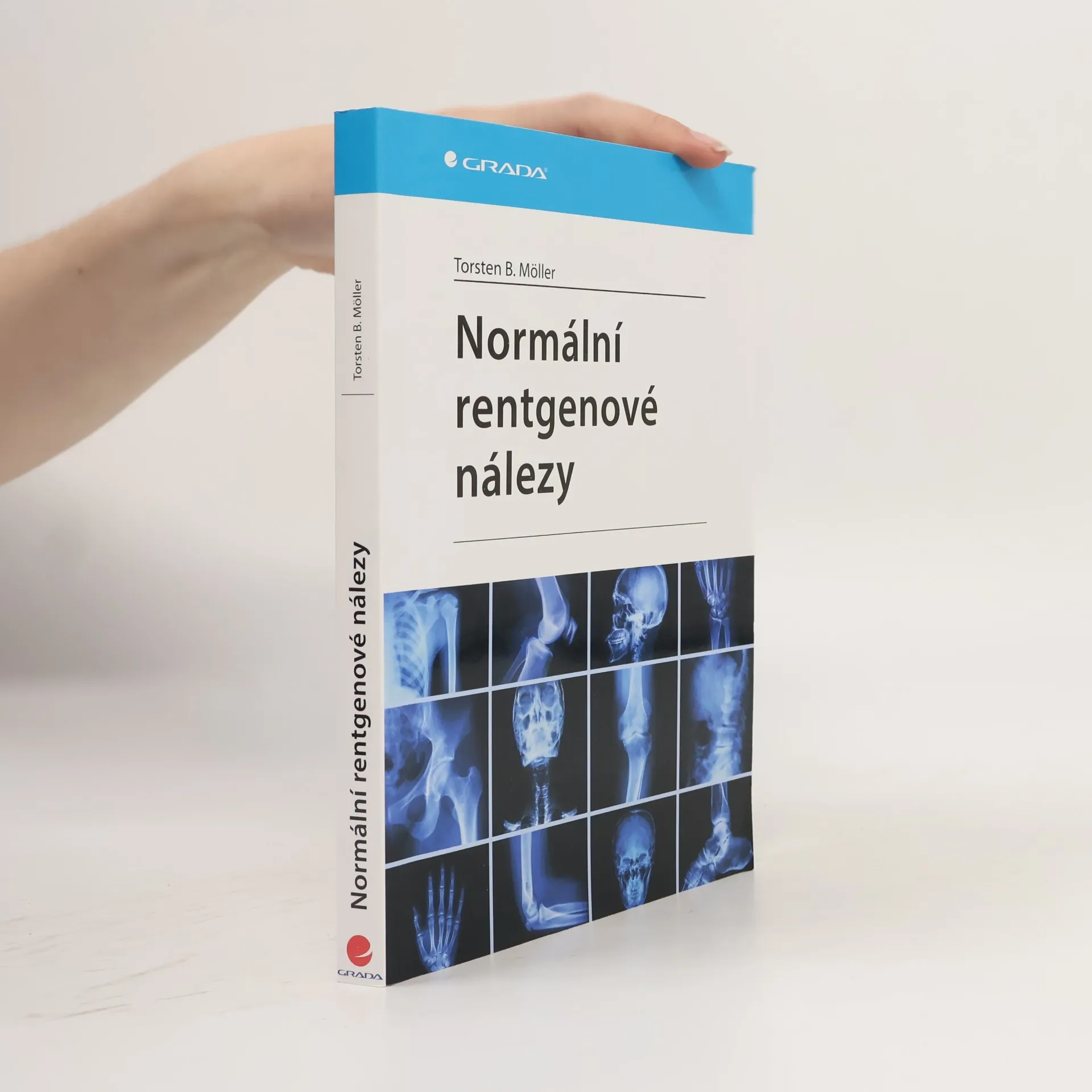
Röntgennormalbefunde
- 259 stránek
- 10 hodin čtení
Ist das normal? Woran kann ich das erkennen? Wie kann ich das objektivieren? Hier finden Sie Antworten: - klassische Normalbefunde aller Röntgenaufnahmetechniken einschließlich KM-Untersuchungen in hoher Abbildungsqualität - direkt in die Aufnahmen eingezeichnet sind wichtige Daten (Maße, Winkel und andere Kriterien des Normalen) - übersichtliche Checklisten zur Systematik der Bildbetrachtung - hilfreiche Befundformulierungen - mit „digitalem Röntgen“ und neueren Kontrastmittel-Darstellungen
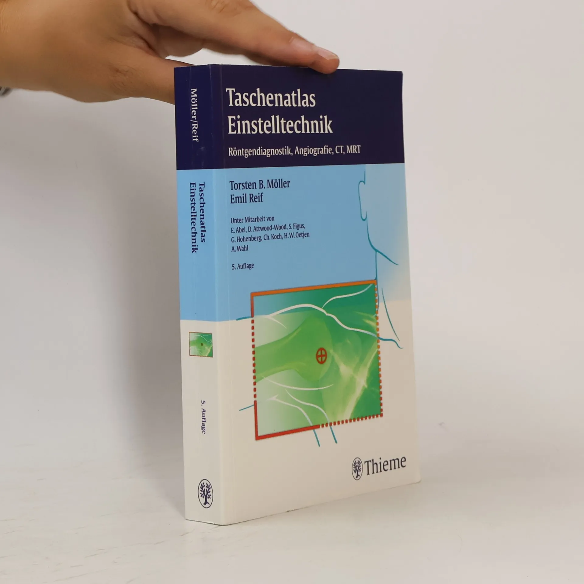
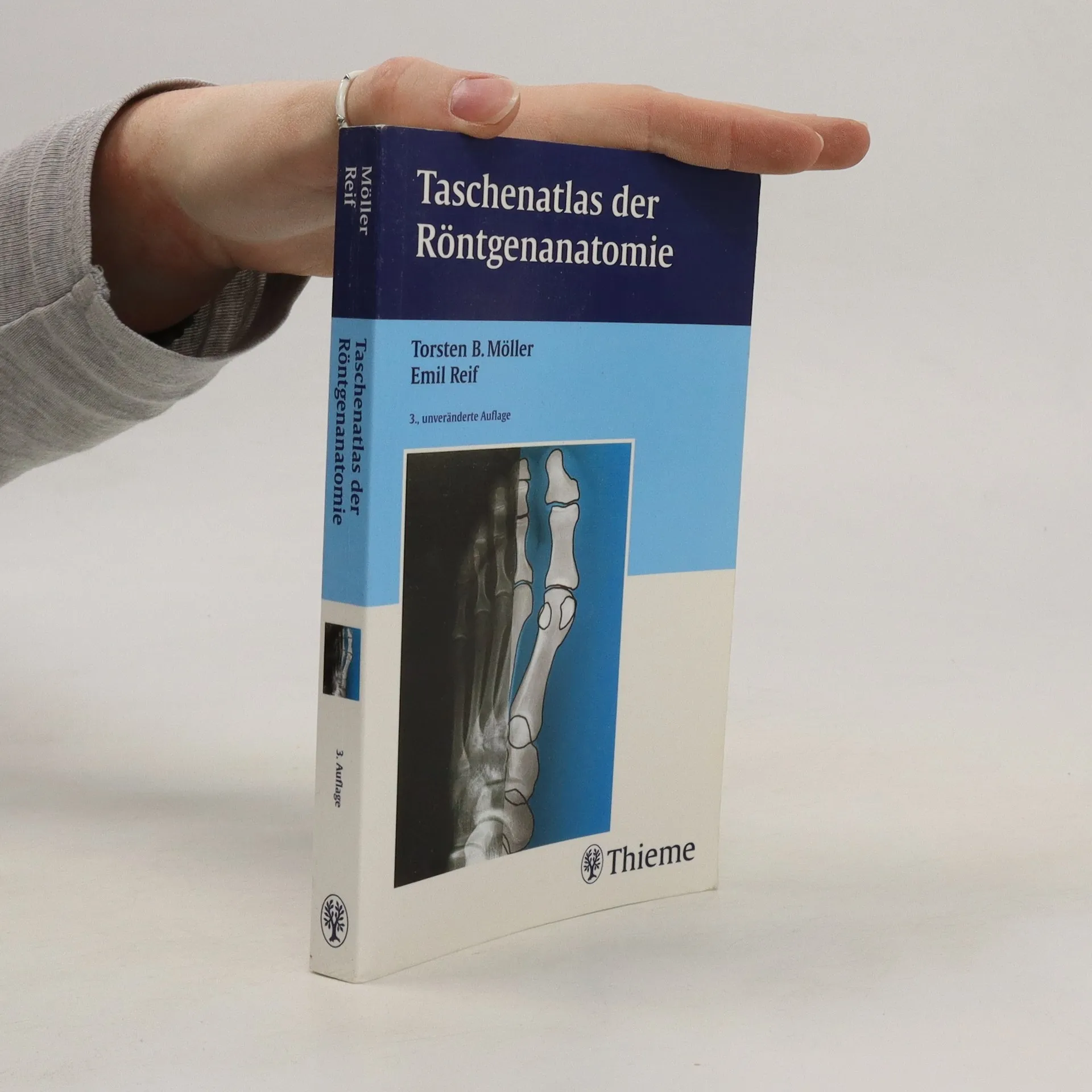
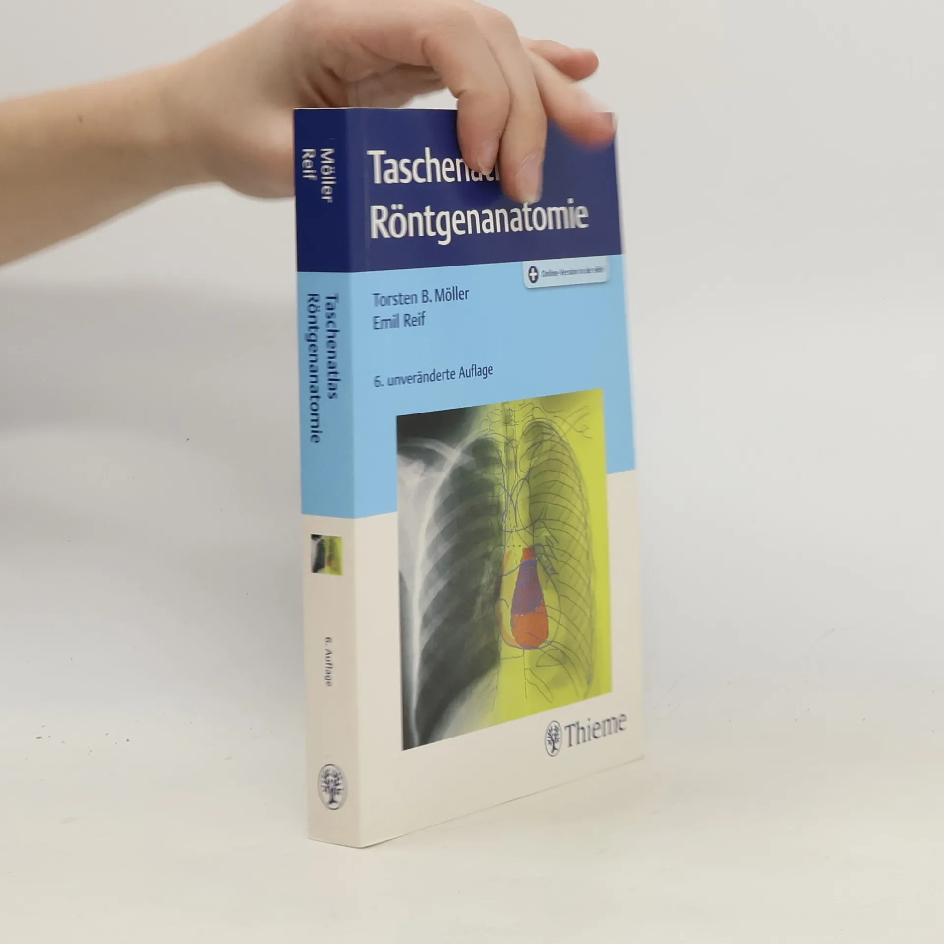
Taschenatlas Röntgenanatomie
- 391 stránek
- 14 hodin čtení
Präzise im Detail: Alle gängigen Standard- und Spezialeinstellungen der konventionellen Radiologie einschließlich aller Kontrastmitteluntersuchungen werden beschrieben. Extrem anschaulich: Alle klinisch relevanten anatomischen Strukturen werden in einer dem Röntgenbild direkt gegenübergestellten Zeichnung wiedergegeben und benannt. Immer griffbereit, handlich und übersichtlich: Das praktische Taschenbuchformat macht es leicht, das Buch immer dabei zu haben. Der einfache Aufbau erspart mühsames Suchen in Inhalts- oder Sachverzeichnissen.
Dieses Buch vermittelt Ihnen die Einstelltechnik in der Röntgendiagnostik. Entwickelt unter Mitarbeit von MTRA und Lehr-MTRA aus verschiedenen Instituten entstand ein Buchkonzept, das sich in der Praxis bewährt hat. Lehr- und Arbeitsbuch für alle Fragen derkorrekten Durchführung einer Röntgenuntersuchung, nicht nur für MTRA, sondern auch für den anordnenden Arzt. Dieses Werk setzt Maßstäbe und definiert den Standard, den Sie für eine gute Aufnahmebenötigen. In kurzer und prägnanter Form erfahren Sie alle nötigen Details zur Aufnahmetechnik, Patientenvorbereitung, Lagerung und Einstellung. Die Kombination von Schemazeichnungen mit qualitativhochwertigen Original-Röntgenaufnahmen schult Ihr Auge unddemonstriert Ihnen durch die "Befund-im-Bild-Darstellung" die wesentlichen Kriterien, die eine gute Aufnahme von einer schlechtenunterscheiden. Anmerkungen zu Varianten und regelmäßigeTipps und Tricks machen dieses Buch zu einem unverzi chtbaren Handwerkszeug für die tägliche Praxis
MR-Atlas des muskuloskelettalen Systems
- 308 stránek
- 11 hodin čtení
Taschenatlas der Röntgenanatomie
- 404 stránek
- 15 hodin čtení
Eine ideale Taschenreferenz zur Röntgenanatomie, die alle Standard- und Spezialeinstellungen der Radiologie sowie Kontrastmitteluntersuchungen beschreibt. Klinisch relevante anatomische Strukturen werden detailliert dargestellt und benannt. Das handliche Format ermöglicht eine einfache Handhabung und schnellen Zugriff auf Informationen.
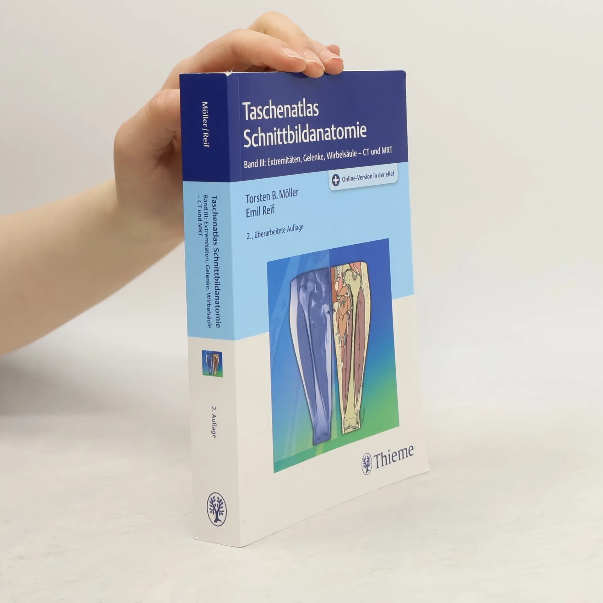
Taschenatlas Schnittbildanatomie 03
Extremitäten, Gelenke, Wirbelsäule - CT und MRT - 2., überarbeitete Auflage
- 480 stránek
- 17 hodin čtení
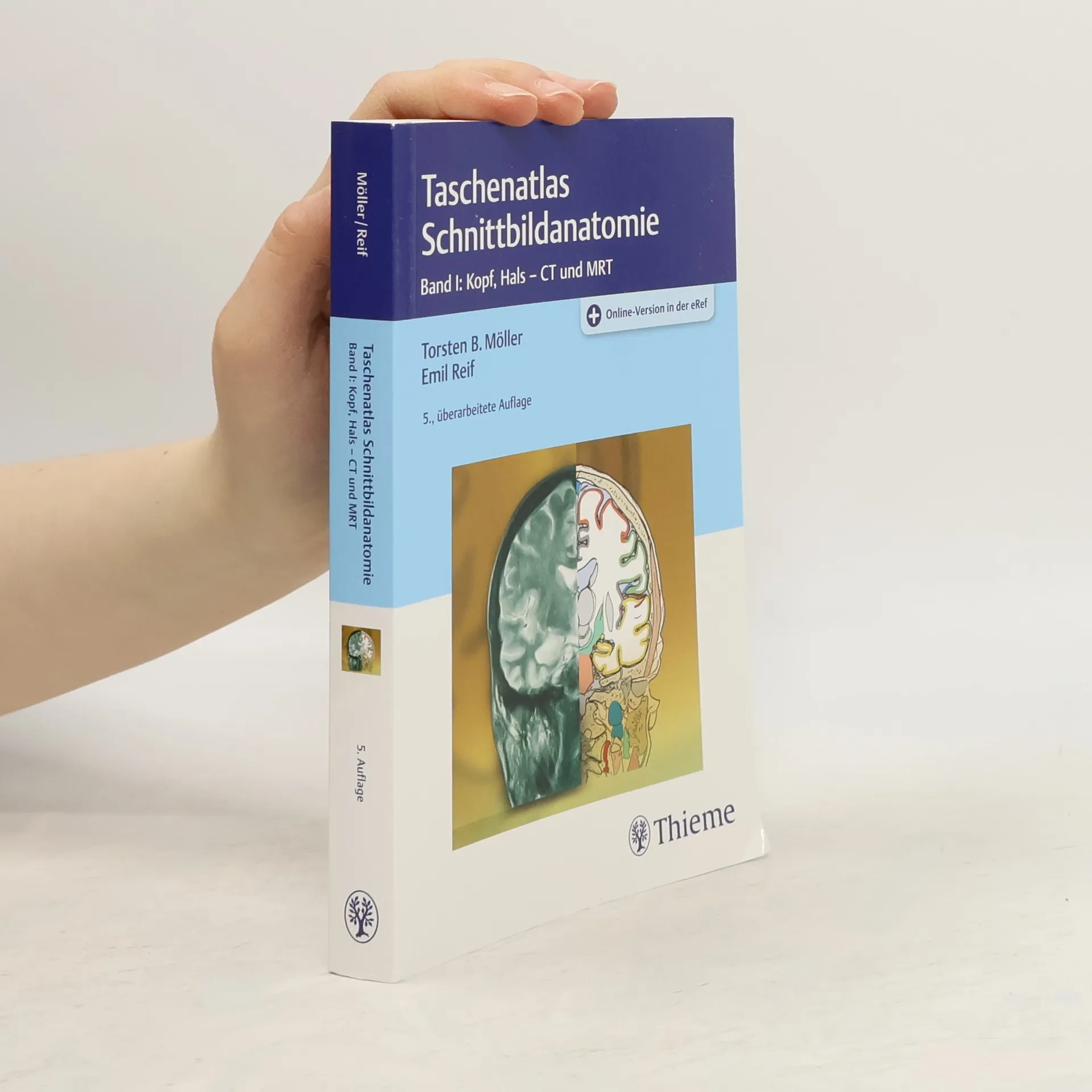
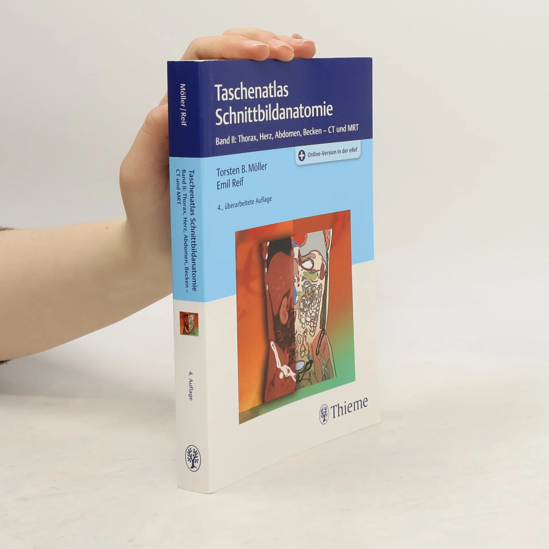
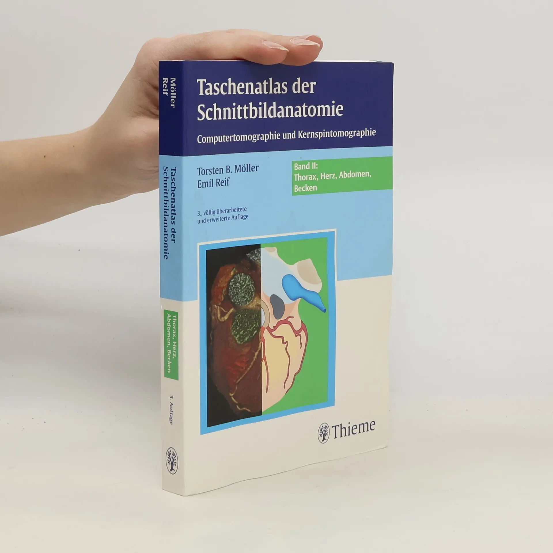
Taschenatlas der Schnittbildanatomie
Thorax, Herz, Abdomen, Becken - 3., völlig überarbeitete und erweiterte Auflage
- 331 stránek
- 12 hodin čtení
Detailgenauigkeit in Perfektion und Farbe Das Pathologische vom Normalen Für die Erstellung und Interpretation von MRT- und CT-Aufnahmen sind Kenntnisse der Schnittbildanatomie unerlässlich. Im eingängigen Konzept stellen Torsten Möller und sein Team jeder Schnittbildaufnahme eine deckungsgleiche Zeichnung gegenüber, auf der die anatomischen Strukturen detailliert eingezeichnet sind. Durch das ausgeklügelte Farbsystem werden alle Strukturen klar zugeordnet. Auß - Scoutzeichnungen zeigen die genau Lage des Schnitts - Beschriftungen direkt am Bild - mit Bildern aus der neuesten Gerätegeneration - in der Linien und Beschriftungen können aus- und eingeblendet werden Jederzeit Der Inhalt des Buches steht Ihnen ohne weitere Kosten digital in der Wissensplattform eRef zur Verfügung (Zugangscode im Buch). Mit der kostenlosen eRef App haben Sie zahlreiche Inhalte auch offline immer griffbereit.
Taschenatlas Röntgenanatomie
Online-Version unter www.thieme.de - 6. unveränderte Auflage
- 400 stránek
- 14 hodin čtení
Röntgennormalbefunde - 5. Auflage
- 270 stránek
- 10 hodin čtení
Ist das normal? Woran kann ich das erkennen? Wie kann ich das objektivieren? Hier finden Sie Antworten: - Klassische Normalbefunde aller Röntgenaufnahmetechniken einschließlich KM-Untersuchungen - Wichtige Daten sind direkt in die Aufnahmen eingezeichnet (Maße, Winkel und andere Kriterien des Normalen) - Übersichtliche Checklisten zur Systematik der Bildbetrachtung - Hilfreiche Befundformulierungen - Mit „digitalem Röntgen“ und neueren Kontrastmitteldarstellungen
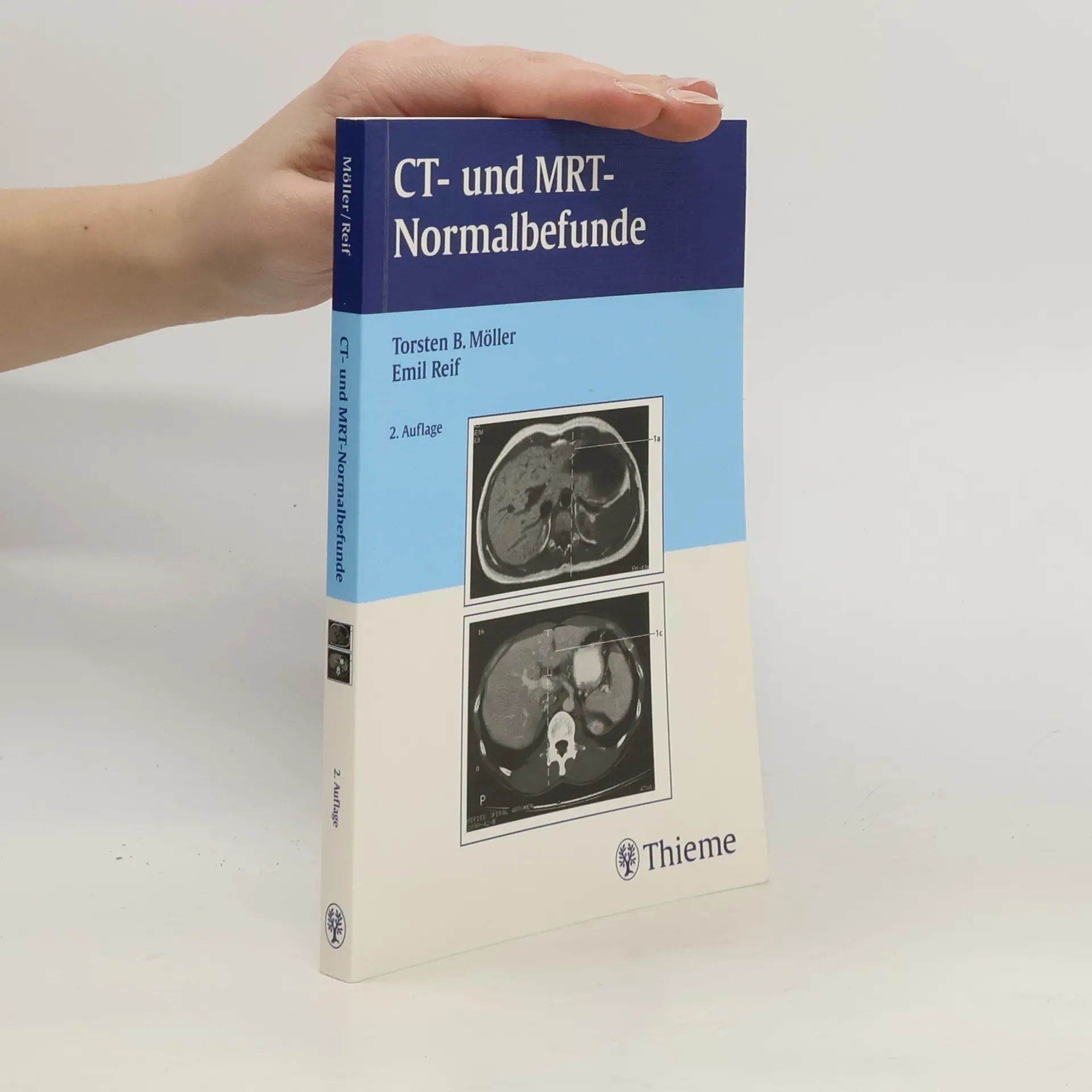
CT- und MRT-Normalbefunde - 2. Auflage
- 250 stránek
- 9 hodin čtení
Der ideale Einstieg in die Schnittbildtechniken: klassische Normalbefunde der gängigen CT- und MRT-Untersuchungen. Direkt in die Aufnahmen eingezeichnet sind Maße, Winkel und andere Kriterien des Normalen. Erlernen Sie die Systematik der Bildbetrachtung: Wie schaue ich mir ein Bild an? Welche Strukturen betrachte ich in welcher Reihenfolge? Worauf muss ich dabei besonders achten?